What is bronchoscopy?
Bronchoscopy is a procedure in which a thin, flexible tube carrying a camera and a light is passed through the nose or mouth, past the vocal cords, and into the windpipe and the branching airways of both lungs. It allows the pulmonologist to see the airway lining directly, to take samples of fluid, cells and tissue, and to carry out treatments inside the airway without any surgical incision.
Two instruments are used. The flexible bronchoscope, performed under local anaesthesia with conscious sedation, is the workhorse for diagnosis and reaches far into the smaller airways. The rigid bronchoscope, performed under general anaesthesia in the operating theatre, provides a wide working channel and airway control, and is used for major therapeutic work such as removing a large foreign body, controlling bleeding, or placing an airway stent.
Bronchoscopy answers questions that scans cannot. A CT can show a shadow, a narrowing or an enlarged node, but only tissue and microbiology can say whether it is cancer, tuberculosis, fungal infection or inflammation, and that distinction determines the entire treatment plan.
Symptoms and findings that lead to bronchoscopy
- Coughing up blood, whether streaks or larger volumes, with no clear explanation.
- Persistent cough lasting more than six to eight weeks that has not responded to treatment and has no diagnosis.
- An abnormal chest X-ray or CT: a lung mass or nodule, persistent collapse of a lobe, a non-resolving patch of pneumonia, cavities, or enlarged lymph nodes.
- Suspected lung cancer needing tissue for diagnosis, molecular testing and staging.
- Suspected tuberculosis or fungal infection when the patient cannot produce sputum or when sputum tests are repeatedly negative.
- Pneumonia in an immunosuppressed patient, where identifying the exact organism quickly changes treatment.
- Localised wheeze or stridor, suggesting narrowing of a large airway.
- Suspected inhaled foreign body, particularly in children and elderly patients.
- Interstitial lung disease requiring lavage or lung biopsy to reach a diagnosis.
Diagnostic techniques through the bronchoscope
| Technique | What it obtains |
|---|---|
| Airway inspection | Direct view of tumours, inflammation, narrowing, bleeding source, secretions and vocal cord movement |
| Bronchial washings and brushings | Cells and microbiological samples from a specific segment |
| Bronchoalveolar lavage | Fluid from the air sacs for infection, cell counts and malignant cells |
| Endobronchial biopsy | Tissue from a visible airway lesion |
| Transbronchial lung biopsy | Small pieces of lung tissue beyond the airway wall, usually under fluoroscopic guidance |
| Transbronchial cryobiopsy | Larger, better preserved tissue samples for interstitial lung disease |
| EBUS-TBNA | Ultrasound-guided needle sampling of lymph nodes and masses next to the airway |
| Radial EBUS and navigation | Access to small peripheral nodules that lie beyond the reach of direct vision |
Samples are sent for cytology and histopathology, for smear, GeneXpert and mycobacterial culture, for bacterial and fungal culture, and, where lung cancer is confirmed, for molecular and PD-L1 testing that determines eligibility for targeted therapy and immunotherapy.
Therapeutic bronchoscopy
Bronchoscopy is not only diagnostic. Therapeutic applications include removal of inhaled foreign bodies such as a peanut, seed, tooth or pin, and suction clearance of thick secretions or mucus plugs causing lobar collapse. Bleeding can be controlled with cold saline, adrenaline, tranexamic acid or endobronchial blockers. Other uses are debulking of airway tumours using cryotherapy, electrocautery, argon plasma coagulation or laser, balloon dilatation of narrowed airways, placement of silicone or metallic stents to relieve airway obstruction, and delivery of valves for selected patients with severe emphysema or persistent air leak. These techniques are described in more detail on the interventional pulmonology page.
Preparation, the procedure and recovery
Before the procedure
You will be asked to fast for about six hours for solids and two hours for clear fluids. Bring a list of all medications. Blood thinners such as clopidogrel, warfarin or newer anticoagulants usually need to be stopped for a defined period, and this must be planned with the prescribing doctor rather than stopped independently. Diabetes medication and insulin doses are adjusted for the fasting period. Blood tests for clotting and blood counts, an ECG and, in some patients, a review of recent imaging are arranged. Tell the team about allergies, asthma, heart disease, previous anaesthesia problems and pregnancy.
During the procedure
Monitoring of heart rate, blood pressure and oxygen saturation is continuous, and supplemental oxygen is given. Local anaesthetic spray or gel numbs the nose and throat, and sedation is given through a vein so that most patients are drowsy and comfortable and remember little afterwards. The scope is passed gently, the airways are inspected systematically, and the planned samples are taken. A typical diagnostic procedure takes twenty to forty-five minutes.
After the procedure
You will be observed until the sedation wears off. Because the throat remains numb, nothing is taken by mouth for about two hours, and a swallow of water is tested before eating. A sore throat, hoarse voice, mild cough and small streaks of blood in the sputum for a day or two are expected. If sedation was used, do not drive, operate machinery or sign legal documents for the rest of the day, and arrange for someone to accompany you home. Most patients return to normal activity the next day. Preliminary findings are discussed on the same day, and microbiology and pathology reports follow over several days.
Risks and safety
Bronchoscopy is a safe procedure in experienced hands, and serious complications are uncommon. Recognised risks include bleeding at a biopsy site, which is usually minor and self-limiting, and pneumothorax (mainly after transbronchial biopsy), which occasionally needs a chest drain. Other risks are transient fever, low oxygen levels during the procedure, airway spasm in patients with asthma, and reactions to sedation or local anaesthetic. The risk profile is discussed and consent is taken before every procedure, and the benefit of a definite diagnosis is weighed against these risks for each patient.
Contact the hospital promptly after discharge if you cough up more than a small amount of blood, develop increasing breathlessness or chest pain, or have a high fever.
Frequently asked questions
Is bronchoscopy painful?
It is not painful. Local anaesthesia numbs the airway and intravenous sedation keeps you drowsy and relaxed. The commonest sensations are a brief urge to cough and pressure in the throat. Most patients recall very little of the procedure afterwards.
Will I be asleep, and is general anaesthesia needed?
Flexible diagnostic bronchoscopy is normally done under conscious sedation rather than general anaesthesia. Rigid bronchoscopy and complex therapeutic procedures are done under general anaesthesia in the operating theatre.
How long does it take and will I be admitted?
Most diagnostic procedures take twenty to forty-five minutes, and patients go home after two to four hours of observation. Admission is reserved for therapeutic procedures, patients with significant comorbidity, or when a complication requires monitoring.
Can I eat before and after?
Fasting is required for about six hours before. Afterwards, wait roughly two hours until the throat sensation returns, then start with sips of water before taking food, to avoid choking while the throat is still numb.
Should I stop my blood thinners?
Usually yes, but only after discussion with the doctor who prescribed them, since stopping them carries its own risks. Aspirin can often be continued for simple procedures, while clopidogrel and anticoagulants generally need to be withheld before biopsy.
When will the results be available?
Visual findings are known immediately. Cytology and GeneXpert results usually take one to three days, histopathology three to seven days, and mycobacterial culture several weeks. The report is interpreted together with the imaging and clinical picture at the follow-up consultation.
Can bronchoscopy be repeated?
Yes. It can be repeated safely when the first procedure is non-diagnostic, when disease progresses, or when treatment such as tumour debulking or secretion clearance needs to be repeated.
Related conditions and procedures
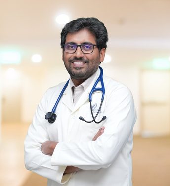
Consult Dr. Gogineni Ratnakara Rao
Apollo Hospitals, Financial District: Monday to Saturday, 8:30 AM to 5:00 PM. Anything in Lung Clinic, Chanda Nagar: Monday to Saturday (Thursday closed), 6:00 PM to 9:00 PM.